Healing runs in overlapping stages, and no single week marks the end. New bone starts forming during the first week.12 Cleveland Clinic puts the first round of healing at about one week and the jawbone's fusion around the implant at three to nine months.3 Stability then dips somewhere in weeks 2 to 4 before it recovers.456 The gum seal around the implant takes 6 to 8 weeks to form.78
When the crown goes on depends on the loading protocol. Cochrane defines immediate loading as within 1 week, early loading as between 1 week and 2 months, and conventional loading as after 2 months.9
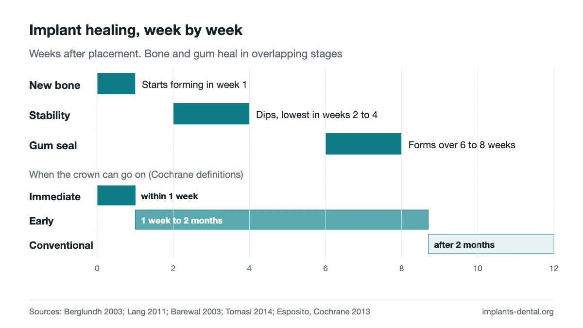
Week 1: what happens around the implant
Drilling leaves a space between the implant and the bone. In animal studies, a blood clot fills it first. Granulation tissue follows and is replaced by a provisional matrix, and bone formation starts during the first week.1 That study used 20 Labrador dogs and 160 experimental devices, so the timings it reports come from animals.
Human biopsies point the same way. In 49 volunteers, new bone formation started during the first week in the trabecular regions and increased gradually up to 42 days.2 The volunteers received small experimental devices (4 mm x 2.8 mm) in the retromolar area, not restorative implants.
Cleveland Clinic says the first round of healing takes about one week, with slight discomfort and swelling, and that you should feel like yourself within a week or so. Over-the-counter pain relievers can help with these symptoms.3 A systematic review of 9 randomized trials found that pain control may matter most during the first 72 hours after placement. NSAIDs significantly reduced postoperative pain and the use of rescue analgesics compared with placebo, and the authors found insufficient evidence to recommend a particular analgesic regimen.10 The American Dental Association says a dentist may recommend soft foods, cold foods and warm soup during healing.11
Weeks 2 to 4: the stability dip
At first an implant is held by its threads cutting into bone. In the dog study, the bone beside the threads, which gives that early grip, was resorbed between 1 and 2 weeks and replaced with newly formed bone. The implants stayed clinically stable throughout, despite the temporary loss of hard tissue contact.1
In people, stability is measured with resonance frequency analysis. One study followed 20 patients with 27 implants. The lowest mean stability came at 3 weeks in every bone type. The drop from baseline to 3 weeks was largest in soft Type 4 bone (8.6%), and so was the rise from 3 to 10 weeks (26.9%). Type 1 and Type 4 bone differed at 3 weeks (P = .004), while Type 1 bone did not change significantly over the 10 weeks. After 5 weeks of healing, the pattern showed no significant difference among bone types.4
Other studies put the low point elsewhere. In 32 patients with 76 early-loaded implants, ISQ dipped significantly and was lowest at week 2, with mean drops of 2.2 ± 3.6 for tissue-level and 2.3 ± 3.7 for bone-level implants.5 A retrospective study of 36 patients and 82 implants found a rise at week 1, a dip at week 4 and a rebound by week 12.6 The measurement schedules differ: the first study did not measure at week 4, and the second did not measure at weeks 2 or 3. Taken together, the studies place the low point somewhere in weeks 2 to 4.
A single stability reading is one check among several. A systematic review of 62 articles found that most reported an overall increase in ISQ over the observation time, with contradictory findings on how RFA relates to marginal bone loss. Its authors consider repeated RFA measurements useful alongside radiographs and other clinical examinations.12
Weeks 6 to 8: the gum seal forms
The gum forms its own seal around the implant. In 21 patients with single-tooth implants and a custom experimental abutment, biopsies were taken at 2, 4, 8 and 12 weeks. At 8 weeks the soft tissue dimension was about 3.6 mm: a barrier epithelium of 1.9 mm and a connective tissue portion of 1.7 mm. Similar dimensions were found at 12 weeks.7
A second biopsy study of 27 implants in 21 patients tracked the inflammation. T and B cells occurred in clusters with a decreasing cell density from 4 to 8 weeks, and the density of vascular structures decreased from 2 to 8 weeks. The authors describe healing of the mucosa as the onset and resolution of inflammation together with increasing tissue attachment to the implant.13
A narrative review concludes that the formation of the biological width and the maturation of the barrier function around transmucosal implants require 6 to 8 weeks of healing. The established soft connective tissue resembles scar tissue in composition, fibre orientation and vasculature.8
Months: when the crown can go on
The familiar wait of 3 to 4 months comes from early Brånemark-era work. A 1981 report covering 2895 implants in the mandible or maxilla described threaded, unalloyed titanium screws that were allowed to heal in situ, without loading, for at least 3 to 4 months.14 That describes the protocol those clinicians used. Cochrane describes the traditional load-free period as 3 to 8 months.9
| Loading protocol | Timing | What studies found | Source |
|---|---|---|---|
| Immediate | Within 1 week | Single crowns, 1 year: OR 0.75 (95% CI 0.32 to 1.76), no significant differences from conventional loading16 | Benic 2014; Cochrane 2013 |
| Early | 1 week to 2 months | Healed-site placement, weighted cumulative survival 98.3% (Type 4B), at least 12 months15 | Gallucci 2018 |
| Conventional | After 2 months | Healed-site placement, weighted cumulative survival 97.7% (Type 4C), at least 12 months15 | Gallucci 2018 |
Cochrane pooled 15 randomized trials comparing immediate with conventional loading. For prosthesis failure in the first year it found RR 1.87 (95% CI 0.70 to 5.01; 8 trials), and for implant failure RR 1.65 (95% CI 0.68 to 3.98; 10 trials). The review covered 26 trials, 1217 participants and 2120 implants, with follow-up of 4 months to 1 year. The mean implant failure rate across all 26 trials was 2.5%, and 3 of the 26 trials had a low risk of bias. The authors found no convincing evidence of a clinically important difference.9 The confidence intervals are wide, so the trials have not shown a clear difference in selected patients. They have not shown that the two are equal.
For single crowns, a meta-analysis of 10 randomized trials found OR 0.75 (95% CI 0.32 to 1.76) for implant survival at 1 year of loading. That conclusion comes mainly from studies of implants inserted with a torque of 20 to 45 Ncm or more, or an ISQ of 60 to 65 or more, and with no need for simultaneous bone augmentation.16
The 2018 ITI-consensus review covered 69 publications on partially edentulous patients with fixed prostheses and rough-surface screw implants 3 to 6 mm in diameter. Weighted cumulative survival across its eight placement and loading protocols ran from 96.0% to 100%, with follow-up of at least 12 months. A pooled analysis was not possible because of considerable heterogeneity, so there are no confidence intervals.15 The ADA notes that some patients wait up to several months for replacement teeth, while others have implants and teeth placed in one visit.11
Upper jaw and lower jaw
One meta-analysis of 21 studies found an early failure rate of 3.14% for maxillary implants and 1.96% for mandibular implants (RR 1.41; 95% CI 1.19 to 1.67; I² = 58%). It identified the anterior maxilla as the most critical site for early loss and the anterior mandible as the one with the best success rate.17 This is a failure rate. It does not measure how fast either jaw heals, and no source reviewed here gives a fixed number of months by jaw.
A review of failure causes lists implant insertion in the maxilla and in the posterior region of the jaws, and in bone of quality types III and IV, among the situations correlated with a higher failure rate. The same review notes that some recent studies suggest modern, moderately rough implants may give similar results whether placed in the maxilla or elsewhere.18 In the stability study above, the softest bone (Type 4) showed the largest dip.4
The oldest numbers are historical. In the Brånemark cohort treated between 1965 and 1980, 81% of maxillary and 91% of mandibular fixtures remained stable (895 fixtures in 130 jaws, followed for 5 to 9 years). Those were machined implants, and the figures are not current success rates.19
What can slow healing
Insertion torque is one measurable factor. In the 36-patient cohort, an insertion torque of 35 Ncm or more was consistently associated with higher ISQ values at all time points.6 The single-crown evidence for same-day loading rests on a torque of 20 to 45 Ncm or more or an ISQ of 60 to 65 or more.16
The failure-cause review also lists a low insertion torque for implants planned for immediate or early loading, heavy smokers, small bone volumes, lack of initial implant stability and shorter implants. It adds that modern, moderately rough implants may give similar results in smokers and with short implants.18 Cleveland Clinic says smoking or vaping can disrupt the healing process.3 The ADA lists chronic illnesses such as diabetes or leukemia as conditions that may interfere with healing after surgery.11
Questions patients ask
How long does osseointegration take?
Cleveland Clinic gives a range of three to nine months for the jawbone to fuse around the implant.3 The 3 to 4 month figure many pages repeat is the unloaded healing period from a 1981 protocol.14 In human biopsies of small experimental devices, bone-to-implant contact was 12.2% to 14.8% at 2 weeks, 32.4% to 48.3% at 4 weeks and 61.6% vs 61.5% at 42 days for the two surfaces tested.2 No source reviewed here defines a completion week. Osseointegration is secured through continuous remodeling and adaptation to function.1
Can I get teeth the same day?
Some patients can. The ADA says others wait up to several months, while some have implants and replacement teeth placed in one visit.11 For single crowns, the 1-year finding of OR 0.75 (95% CI 0.32 to 1.76) comes mainly from implants inserted with a torque of 20 to 45 Ncm or more, or an ISQ of 60 to 65 or more, and with no simultaneous bone augmentation.16 The procedure is covered in same-day implants.
References
- Berglundh T, Abrahamsson I, Lang NP, Lindhe J. De novo alveolar bone formation adjacent to endosseous implants. Clinical Oral Implants Research. 2003;14(3):251–262. doi:10.1034/j.1600-0501.2003.00972.x. PMID 12755774
- Lang NP, Salvi GE, Huynh-Ba G, Ivanovski S, Donos N, Bosshardt DD. Early osseointegration to hydrophilic and hydrophobic implant surfaces in humans. Clinical Oral Implants Research. 2011;22(4):349–356. doi:10.1111/j.1600-0501.2011.02172.x. PMID 21561476
- Cleveland Clinic. Dental Implants. 2026. https://my.clevelandclinic.org/health/treatments/10903-dental-implants. Accessed October 2026.
- Barewal RM, Oates TW, Meredith N, Cochran DL. Resonance frequency measurement of implant stability in vivo on implants with a sandblasted and acid-etched surface. International Journal of Oral & Maxillofacial Implants. 2003;18(5):641–651. PMID 14579951
- van Eekeren P, Said C, Tahmaseb A, Wismeijer D. Resonance frequency analysis of thermal acid-etched, hydrophilic implants during first 3 months of healing and osseointegration in an early-loading protocol. International Journal of Oral & Maxillofacial Implants. 2015;30(4):843–850. doi:10.11607/jomi.3985. PMID 26252037
- Tan S, Bayrakçi N, Gerçek AO. Time-dependent changes in implant stability and the influence of clinical parameters: a 12-week resonance frequency analysis-based study. Journal of Stomatology, Oral and Maxillofacial Surgery. 2026;127(1):102548. doi:10.1016/j.jormas.2025.102548. PMID 40947098
- Tomasi C, Tessarolo F, Caola I, Wennström J, Nollo G, Berglundh T. Morphogenesis of peri-implant mucosa revisited: an experimental study in humans. Clinical Oral Implants Research. 2014;25(9):997–1003. doi:10.1111/clr.12223. PMID 23799997
- Sculean A, Gruber R, Bosshardt DD. Soft tissue wound healing around teeth and dental implants. Journal of Clinical Periodontology. 2014;41(Suppl 15):S6–S22. doi:10.1111/jcpe.12206. PMID 24641001
- Esposito M, Grusovin MG, Maghaireh H, Worthington HV. Interventions for replacing missing teeth: different times for loading dental implants. Cochrane Database of Systematic Reviews. 2013;(3):CD003878. doi:10.1002/14651858.CD003878.pub5. PMID 23543525
- Khouly I, Braun RS, Ordway M, Alrajhi M, Fatima S, Kiran B, Veitz-Keenan A. Post-operative pain management in dental implant surgery: a systematic review and meta-analysis of randomized clinical trials. Clinical Oral Investigations. 2021;25(5):2511–2536. doi:10.1007/s00784-021-03859-y. PMID 33839939
- American Dental Association, MouthHealthy. Implants. https://www.mouthhealthy.org/all-topics-a-z/implants. Accessed October 2026.
- Chen MH, Lyons KM, Tawse-Smith A, Ma S. Clinical significance of the use of resonance frequency analysis in assessing implant stability: a systematic review. International Journal of Prosthodontics. 2019;32(1):51–58. doi:10.11607/ijp.6048. PMID 30677112
- Tomasi C, Tessarolo F, Caola I, Piccoli F, Wennström JL, Nollo G, Berglundh T. Early healing of peri-implant mucosa in man. Journal of Clinical Periodontology. 2016;43(10):816–824. doi:10.1111/jcpe.12591. PMID 27329966
- Albrektsson T, Brånemark PI, Hansson HA, Lindström J. Osseointegrated titanium implants. Requirements for ensuring a long-lasting, direct bone-to-implant anchorage in man. Acta Orthopaedica Scandinavica. 1981;52(2):155–170. doi:10.3109/17453678108991776. PMID 7246093
- Gallucci GO, Hamilton A, Zhou W, Buser D, Chen S. Implant placement and loading protocols in partially edentulous patients: a systematic review. Clinical Oral Implants Research. 2018;29(Suppl 16):106–134. doi:10.1111/clr.13276. PMID 30328194
- Benic GI, Mir-Mari J, Hämmerle CH. Loading protocols for single-implant crowns: a systematic review and meta-analysis. International Journal of Oral & Maxillofacial Implants. 2014;29(Suppl):222–238. doi:10.11607/jomi.2014suppl.g4.1. PMID 24660200
- Fouda AAH. The impact of the alveolar bone sites on early implant failure: a systematic review with meta-analysis. Journal of the Korean Association of Oral and Maxillofacial Surgeons. 2020;46(3):162–173. doi:10.5125/jkaoms.2020.46.3.162. PMID 32606277
- Chrcanovic BR, Albrektsson T, Wennerberg A. Reasons for failures of oral implants. Journal of Oral Rehabilitation. 2014;41(6):443–476. doi:10.1111/joor.12157. PMID 24612346
- Adell R, Lekholm U, Rockler B, Brånemark PI. A 15-year study of osseointegrated implants in the treatment of the edentulous jaw. International Journal of Oral Surgery. 1981;10(6):387–416. doi:10.1016/s0300-9785(81)80077-4. PMID 6809663
This page describes published clinical practice. It is not a treatment plan, and it does not replace an examination.